CytoScanner
Problem
Traditionally, experimental work comprises a sequence of cell preparation, incubation, recording measurements, data processing and finally the evaluation. This process is usually highly repetitive and time consuming. Also, each measurement opposes a disturbance to the cells as they are removed from the incubator. The time required for the measurement can sometimes be longer than one hour. Additionally, researchers continuously have to decide whether they want to spend the night in the lab to make additional measurements. Many of these processes can already be automated, although for a hefty price tag.
Solution
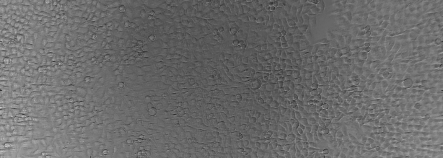
We have developed the CytoScanner platform, a combination of a fully autonomous microscope that can be placed inside the incubator and a software, that enables researchers to easily define custom recording schedules in their office which are then automatically executed by the CytoScanner.
Hardware
The CytoScanner itself is small (30x26x30 cm) and can be placed in most commercially available incubators. The movement range of the current prototype covers common cell-culture containers like 96-well-plates or 200 ml cell-culture flasks. However, the design of the device makes it easily scalable according to your needs. The optics themselves are based on a 8 MP RGB camera and enable a resolution suitable for automatic analysis of common eucaryotic cell lines. Currently, only bright-field microscopy is used for screening. However, we are already working on an epi-flourescence based variant that will be able to image commonly used fluorophores.
Software
The device is controlled by a micro-controller and connected to the internet. As we have designed the device-software to be an easily accessible API, researchers can either use our remote-control software to interact with the device or integrate the CytoScanner into their already existing analysis scripts. Due to the nature of the device-software, interaction from within commonly available programming languages like MatLab, Python or R is possible. The remote-control software will provide the researcher with an easy-to-use modular toolbox to create workflows. The data can then either be stored locally or automatically transferred to shared drives within the network.
Team
Currently, our team consists of two members which are responsible for the hardware, the software and the experimental validation. Both of us are situated at the BioQuant Heidelberg and belong to the Theoretical Systems Biology group. This results in major locational advantages and enables us to test our product with a diverse group of scientists.

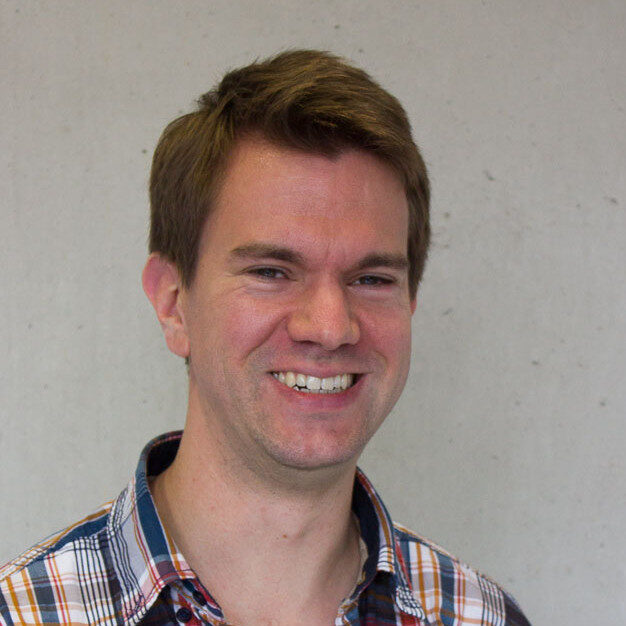
Stefan Kallenberger (Dr. med Dr. rer. nat.) has been doing research in the field of systems biology for several years. His strong background in medicine and physics enables him to approach biomedical research in a very effective manner. Besides working on cancer research by combining mathematical models and live-cell experiments, he likes to develop new technological solutions in life sciences.

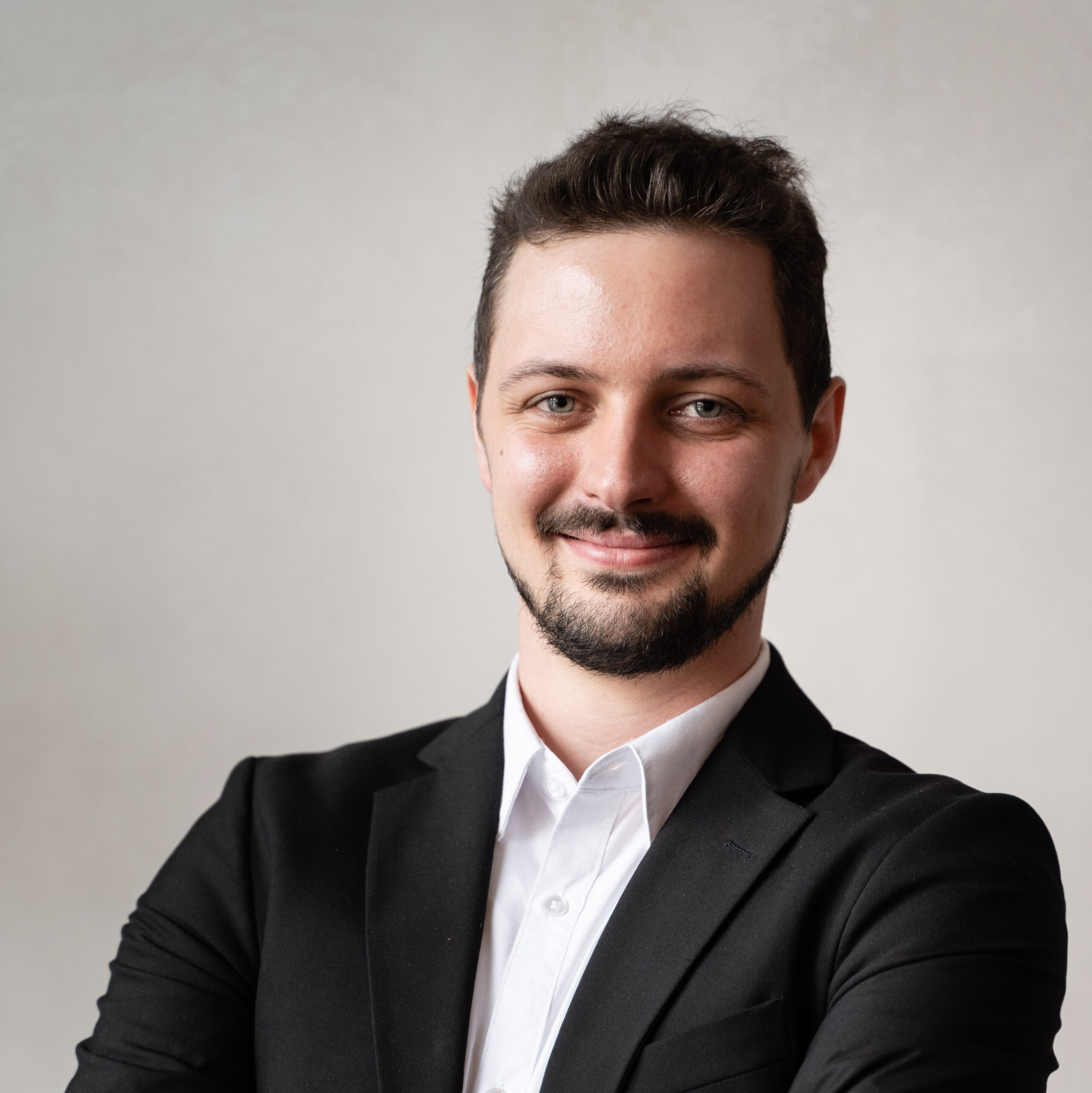
Tim Treis (PhD student) has always been fascinated by the opportunities modern technology offers for biological research. He combines a love for synthetic and systems biology with almost a decade of programming experience. His interest in micro-controllers and 3D printing made it possible to design the custom electronic and optical components within the CytoScanner platform.
If you are interested in working with us during later stages of the project, we’ll be happy to talk about your opportunities. As we are part of an institute associated with Heidelberg University, we’re also interested in talking about possible internships, bachelor or master thesis projects.